
The only tool that is able to allow the development of dermatoscopic analysis, thanks to the possibility, inherent in the technology, of modifying the type of light (polarization and direct light by immersion).
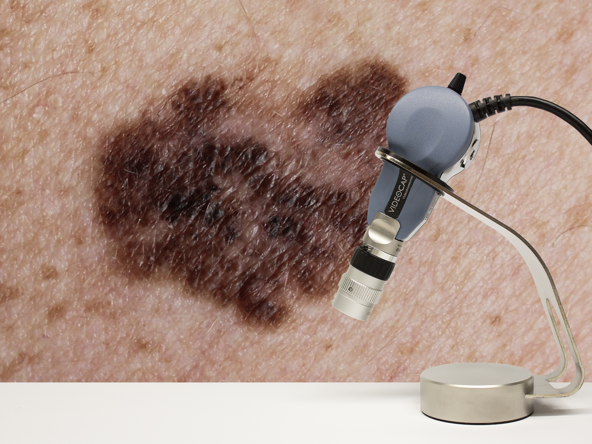
Videodermatoscopy is the field of video diagnostics applied to dermatology, essential for the analysis of moles and the early diagnosis of skin diseases and, therefore, for the prevention of tumors.
Today, it is widely demonstrated how digital videodermatoscopy allows to increase the diagnostic sensitivity by 20-30% compared to simple vision with the naked eye, allowing ever earlier diagnosis of melanoma.
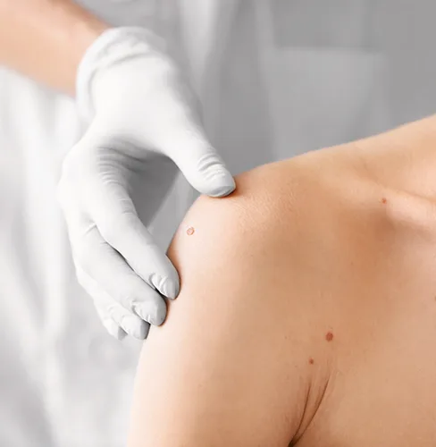
By using videodermatoscopy, the doctor can specifically examine the mole, of which he can accurately determine any alterations or non-homogeneous structures.
Thanks to the information technology support, videodermatoscopy has proceeded with an exponential development and offers, today, bright performances and calculation capacities never seen before.
Thanks to magnifications ranging from 20X to 50X, the analysis of the skin lesion becomes the elective diagnosis in the following pathologies: dermatitis, dermatosis, psoriasis, skin neoplasms, nevi, as well as the entire trichological disease. Videodermatoscopy is also considered very helpful in the following medical disciplines with which the dermatologist must deal: Plastic Surgery, Aesthetic Medicine and Surgery, Angiology/Rheumatology.
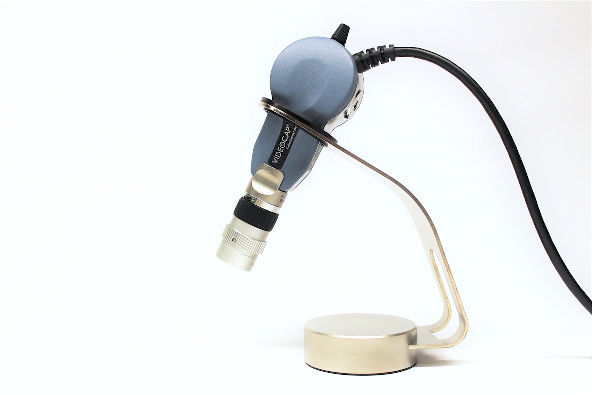
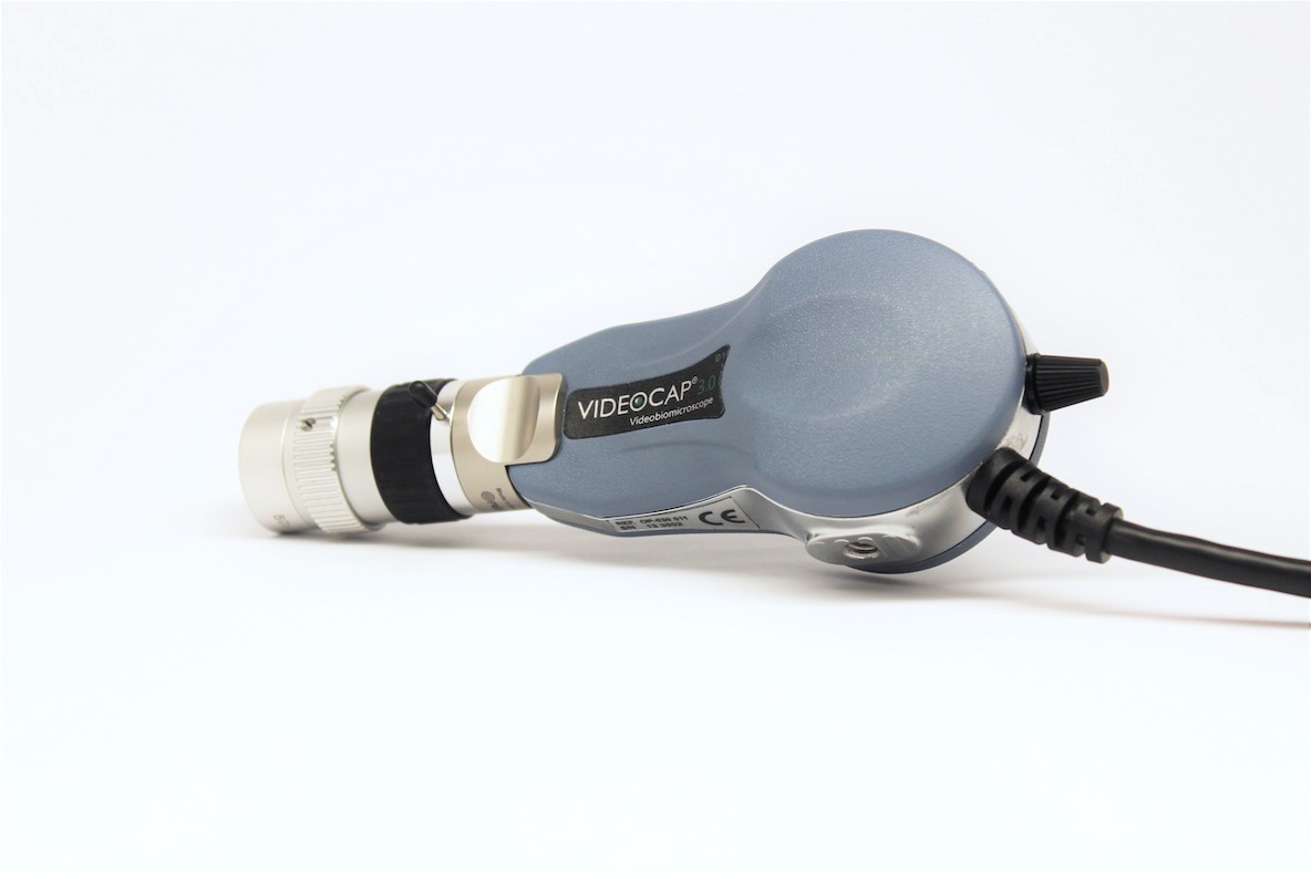
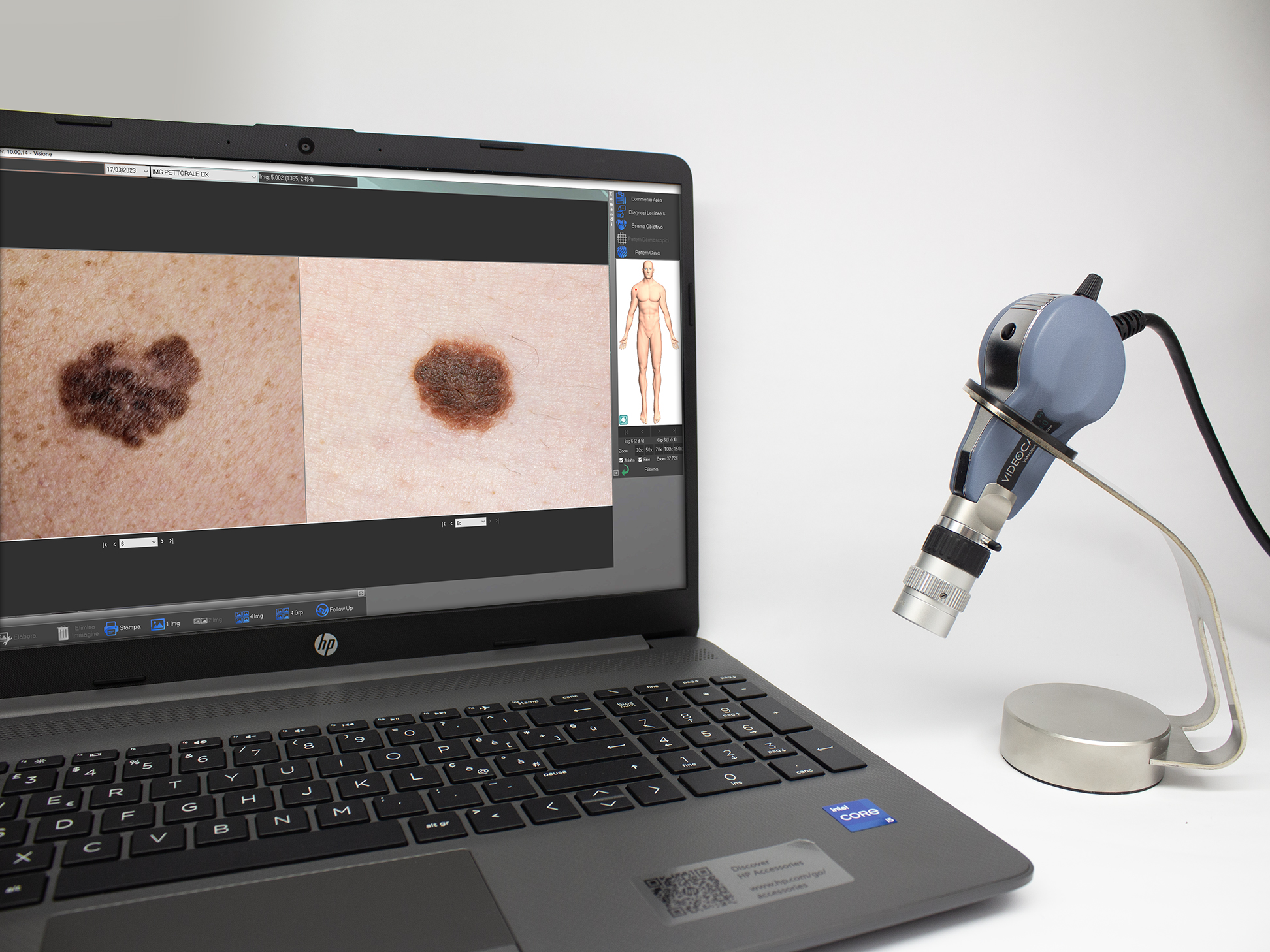
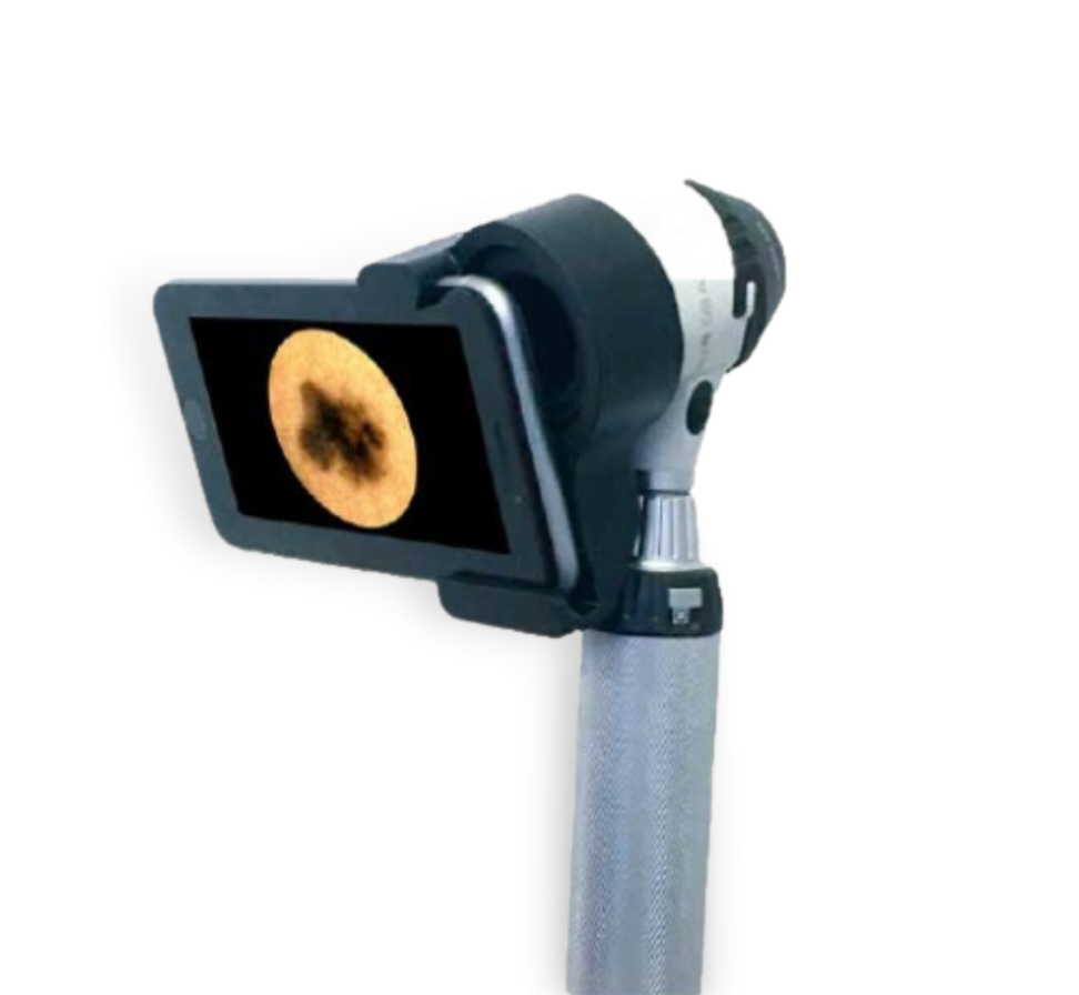
Videocap 3.0® D1 is an epiluminescence, immersion and polarized light videobiomicroscope. It can be used “in contact” and “non-contact” on the basis of the diagnostic needs of the clinician or researcher.
Videocap ® 3.0 D1 connects to a PC via a USB 3.0 interface.
Equipped with on/off button on the back of the probe.
Ergonomic probe with high resolution and chromatic quality, with low weight to reduce wrist fatigue after prolonged periods of work.
Camera sensor: 1/2,5” Rolling; Resolution: 2592H x 1944V pixel (FULL HD)
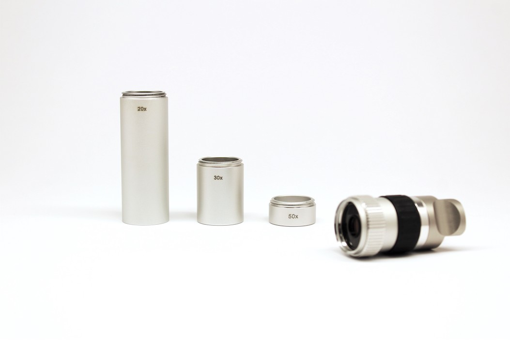
For dermatoscopic analysis it is used a 20-50x lens with specific spacers that allows the visualization of the skin at 20x or 50x and optionally with a 30x spacer. The lens uses a 12 LED system to illuminate the treated part.
With the 20x-50x lens it is possible to acquire images both with white diode light and with polarized light using the metal ring placed on the lens.
There is a manual focus system by using a black adjustment ring placed on the videodermatoscope lens. Fixed magnifications through interchangeable spacers.
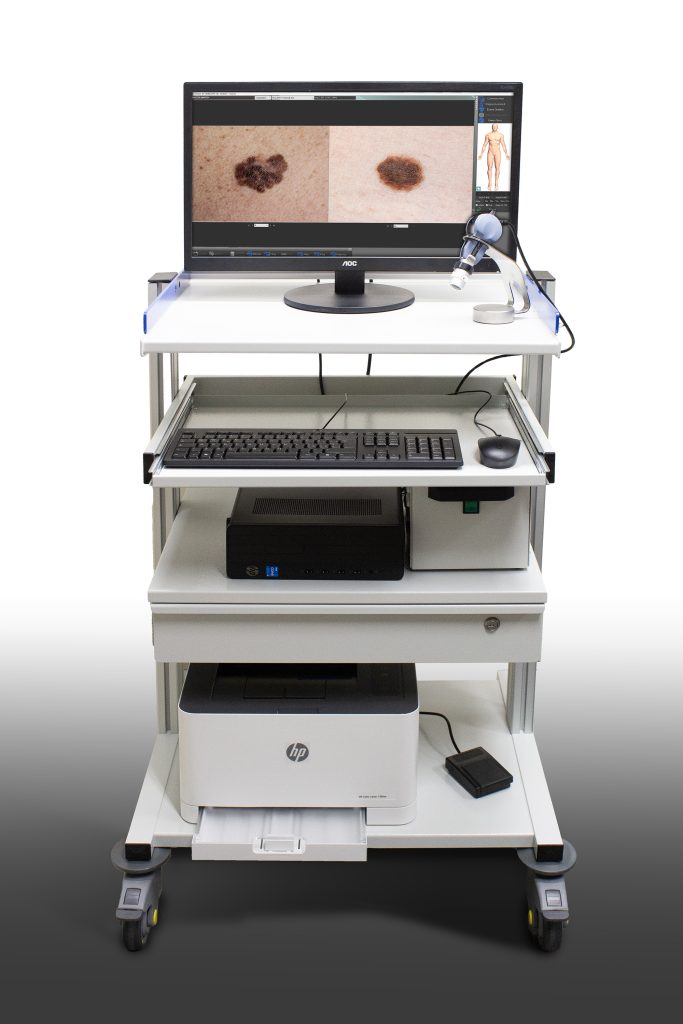
The Videocap ® 3.0 D1 is completed with software for capture and storing images. The Videocap® 3.0 200 REUMA SOFTWARE is part and parcel of the D1 station and is always sold together with the biomicroscope.
Now on the market for more than twenty years Videocap® software, implemented in 32 bit Windows®, has been developed to give the most demanding professionals a tool for differential diagnostics and evaluation of effectiveness in the follow up.
Its reliability and ease of use have made it an irreplaceable work tool.
Videocap® software is designed for transversal use in various medicine disciplines. Thanks to an image storage algorithm, it is able to guarantee high quality photographic image and in vision, without color alteration.
Subject Management, Research, Utilities.
User access in secure and personal mode
It can be installed in the network, to meet the multidisciplinary needs of clinical structrures in order to connect different diagnostic clinics, also thanks to the possibility of using different video imaging devices used in specific specializations (camera, video cameras, histological microscopes, stereomicroscopes, ecotomographs, ultrasounds, videothermographs, etc..).
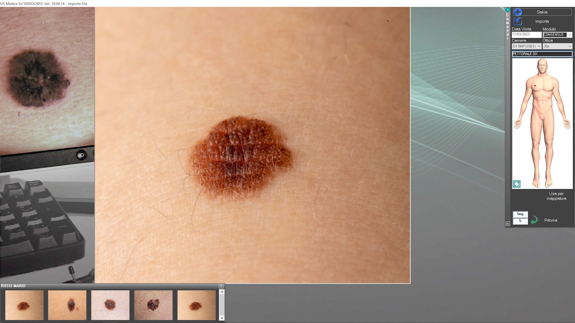
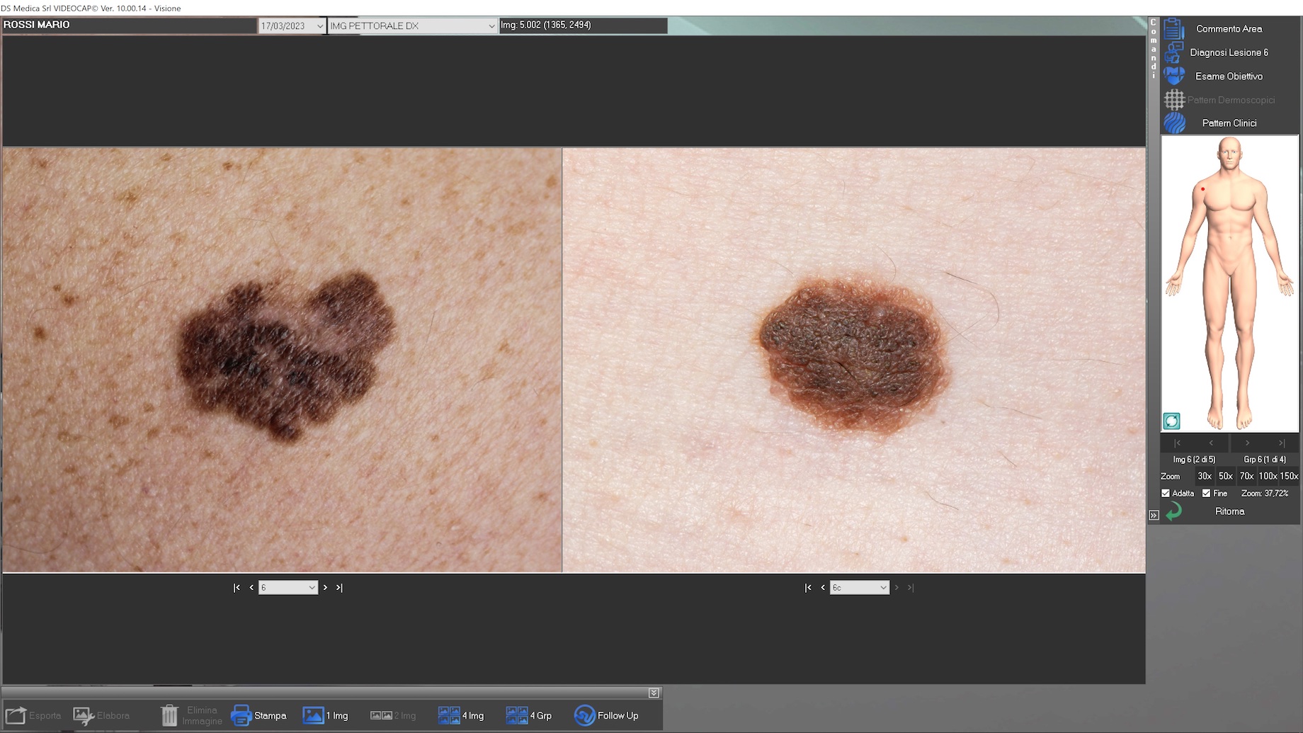
Possibility of recording new visits, examining those already present in the archive, processing the diagnosis and printing the visit report, comparing the images obtained during previous visits.
The search section allows you to research the entire archive, using a system based on keywords.
These can be grouped according to their category (gender, diagnosis, mapping, histological diagnosis, clinical and dermatoscopic patterns, etc.).
Possibility of personalization by the user of the body areas, the various diagnoses, the access account and all that is related to the configuration of use.
Possibility of capture images by positioning the probe on the body area to be examined and focusing on the image. The “in vivo” images will appear on the screen.
The software is enabled for video recording on hard disk or VHS.
Use of the most sophisticated measurement and image analysis tools
Post processing of images for inserting descriptions and geometric figures (arrows, circles, rectangles, freehand selection, grid). Dynamic localization for mapping to real images and bodymaps.
Possibility of comparing the images of a patient obtained during different visits, belonging to different body areas or obtained from other sources (databases, CD-ROM, historical archive created by the user).
Possibility of an accurate and proper follow up.
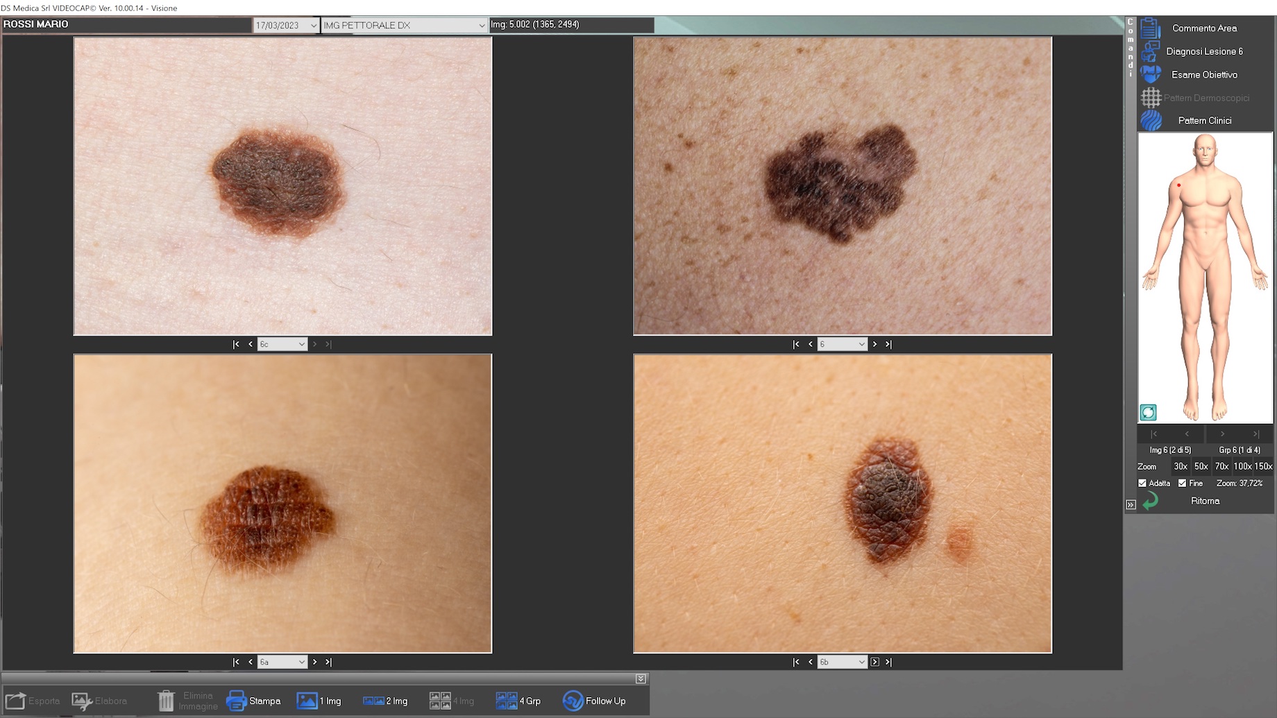
Possibility of correct and complete reporting according to national and international guidelines. Possibility to print different types of reports; 1, 2 and 4-image reports of the same lesion or visit or follow-up to a specific lesion or visit. All reports can be customized according to the user’s needs. Printing of reports with photo map of moles and dermatoscopic images with relative references to the body map.
The software is designed for the use of telemedicine which, however, remains the responsibility of the user.
Videocap® is the DS Medica product line dedicated to epiluminescence, immersion and polarized light video biomicroscopes.
DS Medica S.R.L. | Tax ID/VAT 12676030153
Our customer service to assist in the proper use of software and instrumentation is available Monday through Friday