
Videocapillaroscopes
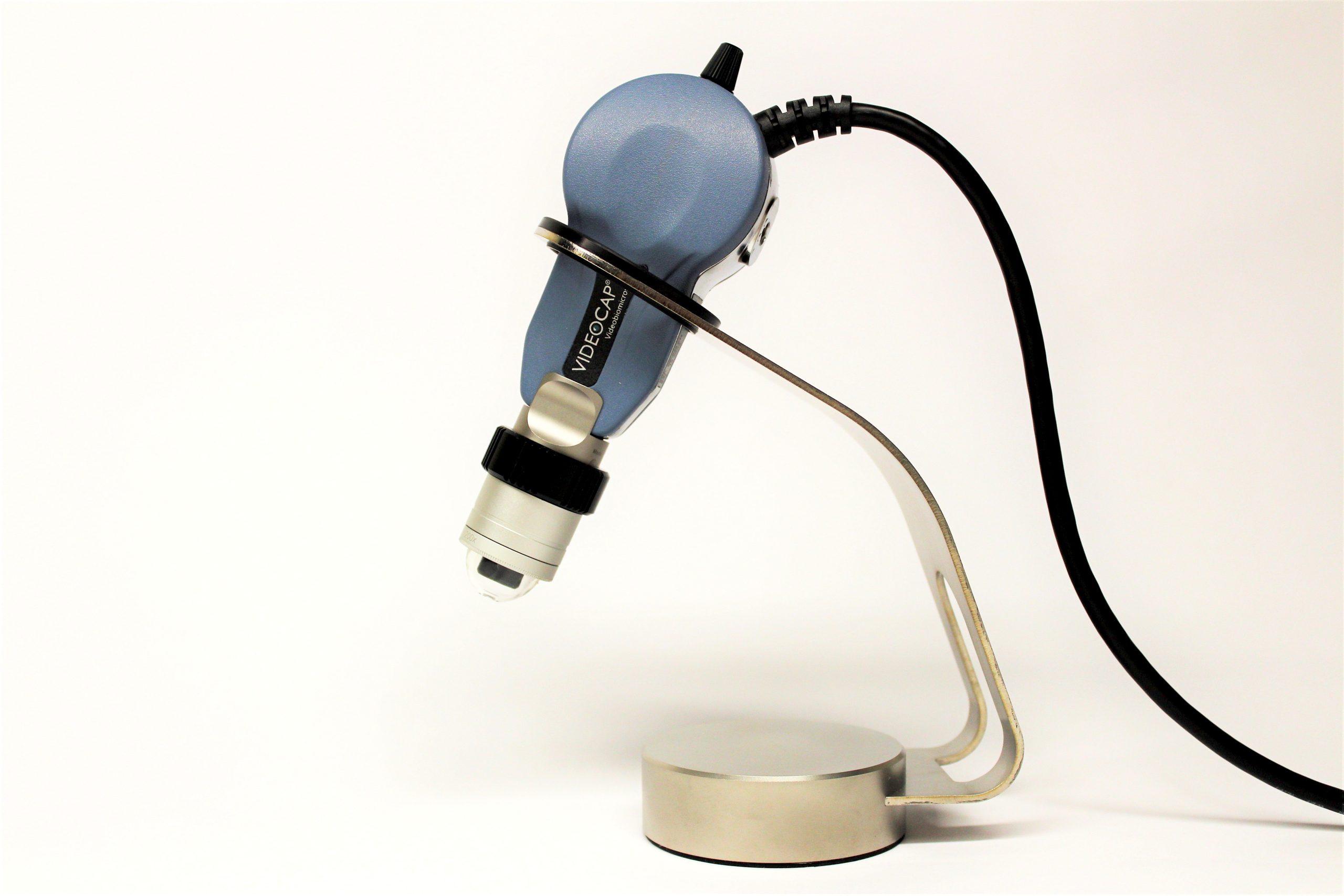
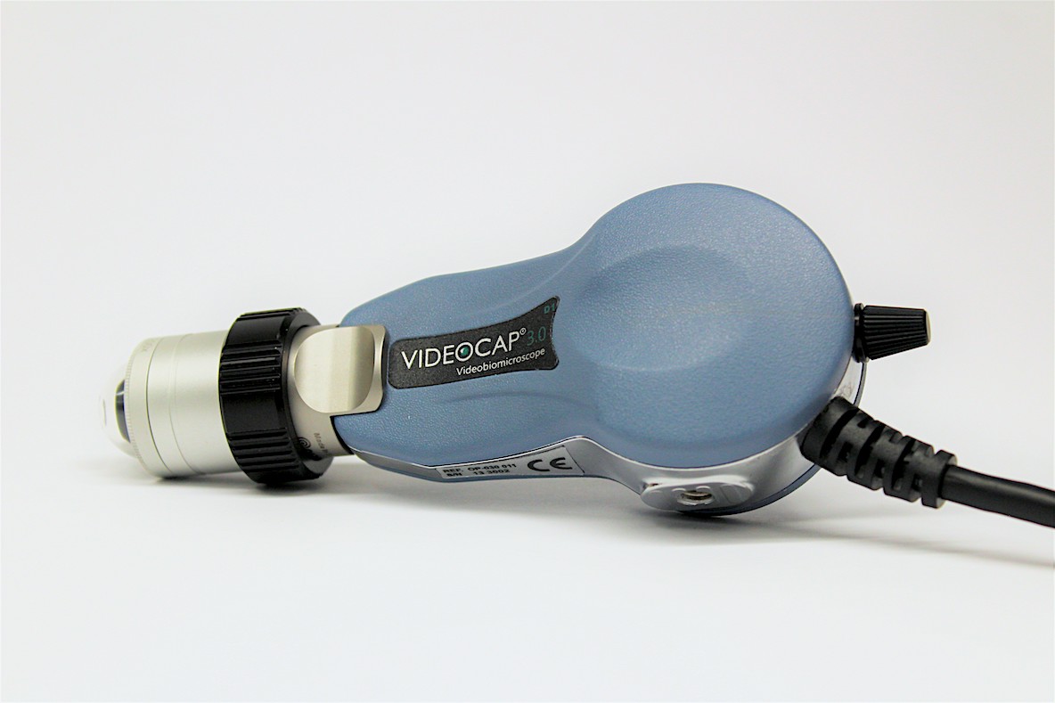
Videocap® 3.0 D1
The only tool that is able to allow the development of capillaroscopic analysis, thanks to the
interchangeable lenses of different magnifications and to the possibility, inherent in the technology, of modifying the type of light (polarization and direct light by immersion).
Capillaroscopy
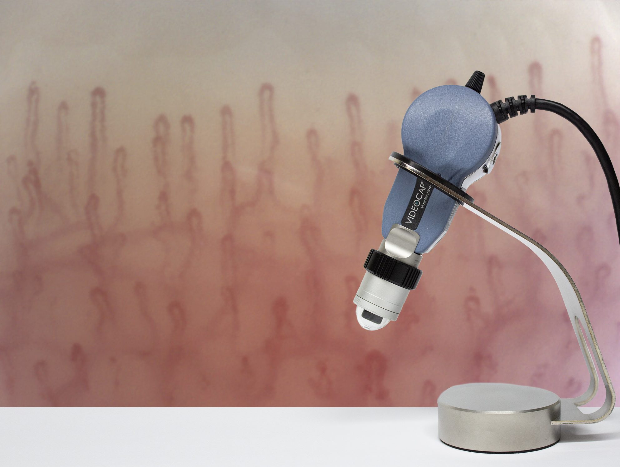
Capillaroscopy is the most widely used technique in the “in vivo” study of microcirculation and in the early diagnosis of systemic sclerosis and related diseases.
The nailfold videocapillaroscopy, in particular, finds elective indication in the study of microangiopathies of rheumatological interest.
In diseases with “scleroderma spectrum disorders” (systemic sclerosis, mixed connective tissue disease, non-differentiated connective tissue disease, pre-scleroderma Raynaud’s phenomenon, dermatomyositis) capillaroscopy is considered a “first level” investigation.
The introduction of video-imaging capillaroscopy has made this investigating technique irreplaceable in the diagnosis of many diseases, as it has allowed to quantify and standardize the colorimetric and morphological parameters.
The device
Videocap®3.0 D1 is the only tool that is able to allow the development of capillaroscopic analysis, thanks to the interchangeable lenses of different magnifications and to the possibility, inherent in the technology, of modifying the type of light (polarization and direct light by immersion).
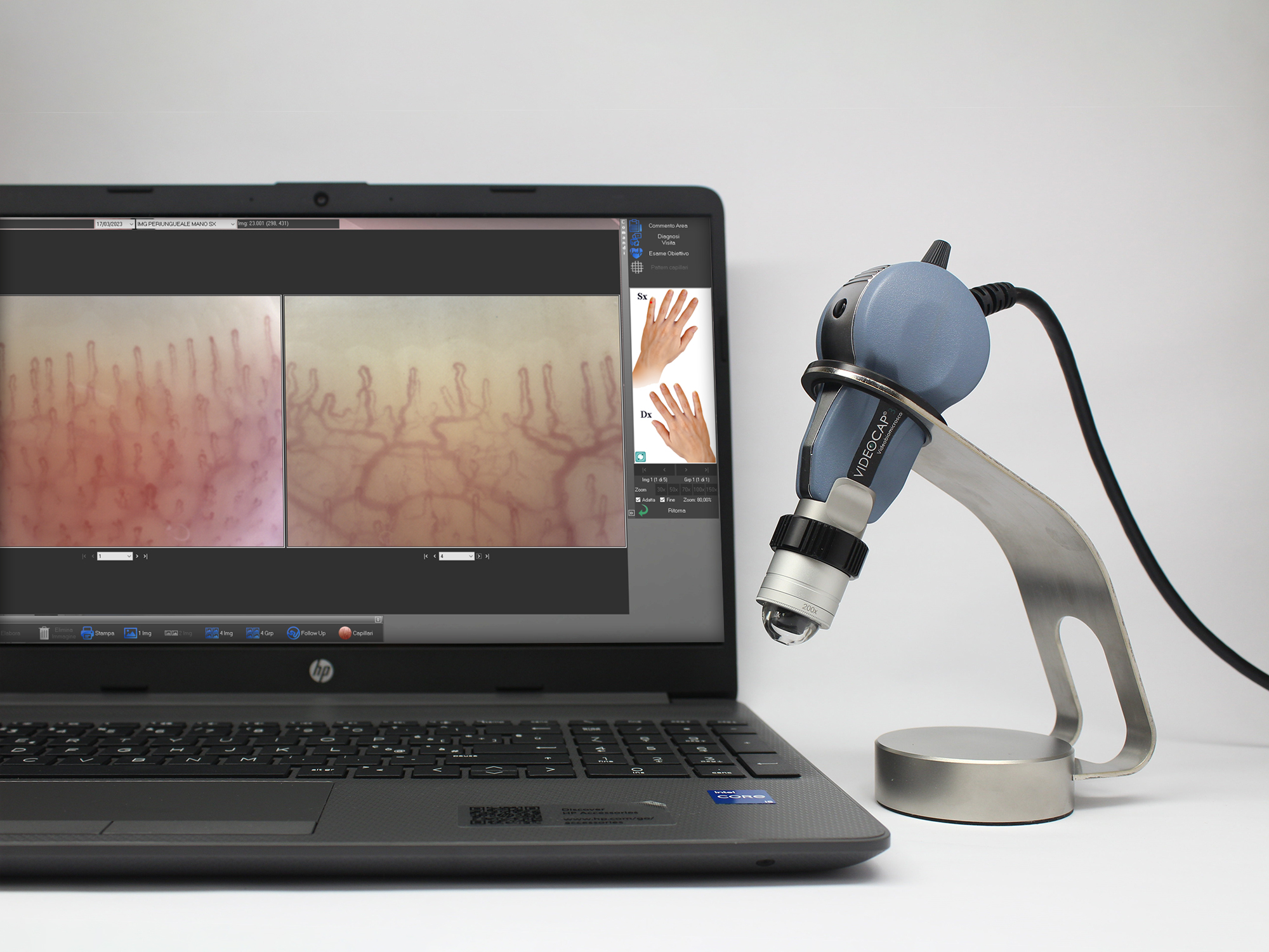
Videocap ® 3.0 D1 connects to a PC via a USB 3.0 interface.
Equipped with on/off button on the back of the probe.
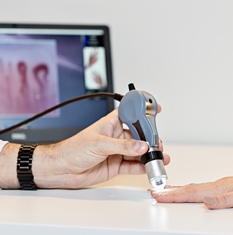
Ergonomic probe with high resolution and chromatic quality, with low weight to reduce wrist fatigue after prolonged periods of work.
Camera sensor: 1/2,5” Rolling; Resolution: 2592H x 1944V pixel (FULL HD)
Main probe features:
- Ergonomics
- Attention to the cleaning and sanitizing operations of all parts of the device
- Easily accessible parts subject to periodic maintenance and cleaning
- Portability
- Practicality and easy to use
Integrated controls:
- White balance
- Brightness regulator
- Contrast
- Image gain factor
- Gamma
- Electronic capture on the image (by a remote footswitch)
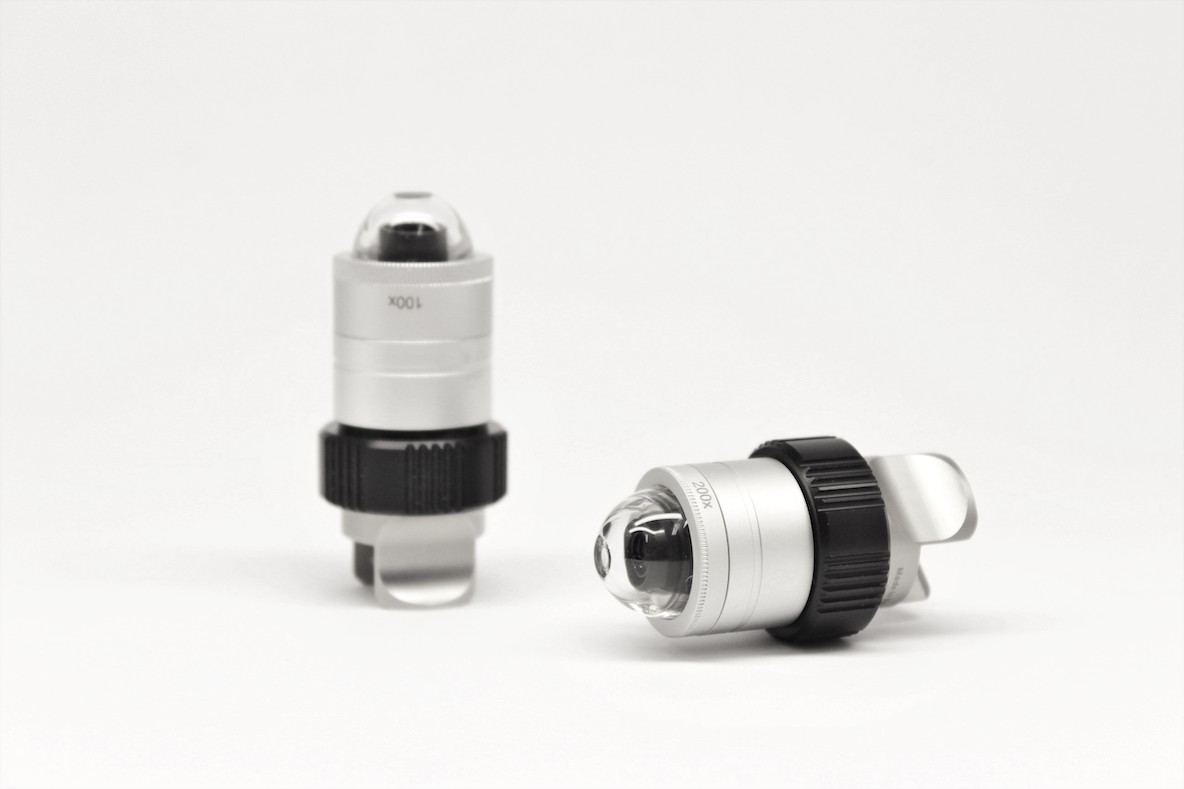
The specific application of the device in rheumatology field is achieved through the use of the 200x lens. The lens uses a 12 LED system to illuminate the treated part. It’s equipped with a plexiglass dome, to protect it against cedar oil.
With the 200x lens it is possible to capture images with cold white diode light using the immersion technique. There is a manual focus system by using a black adjustment ring placed on the videocapillaroscope lens.
Optionaly, is possibile to use a 100x magnification lens.
The software
The Videocap ® 3.0 D1 is completed with software for capture and storing images. The Videocap® 3.0 200 REUMA SOFTWARE is part and parcel of the D1 station and is always sold together with the biomicroscope.
Now on the market for more than twenty years Videocap® software, implemented in 32 bit Windows®, has been developed to give the most demanding professionals a tool for differential diagnostics and evaluation of effectiveness in the follow up.
Its reliability and ease of use have made it an irreplaceable work tool.
Videocap® software is designed for transversal use in various medicine disciplines. Thanks to an image storage algorithm, it is able to guarantee high quality photographic image and in vision, without color alteration.

Peculiarities of the software
Subject Management, Research, Utilities.
User access in secure and personal mode.
It can be installed in the network, to meet the multidisciplinary needs of clinical structrures in order to connect different diagnostic clinics, such as rheumatology, dermatology, radiology, histology, internal medicine, etc., also thanks to the possibility of using different video imaging devices used in specific specializations (camera, video cameras, histological microscopes, stereomicroscopes, ecotomographs, ultrasounds, videothermographs, etc..).
Possibility of recording new visits, examining those already present in the archive, processing the diagnosis and printing the visit report, comparing the images obtained during previous visits.
The search section allows you to research the entire archive, using a system based on keywords.
These can be grouped according to their category (gender, diagnosis, mapping, histological diagnosis, clinical and capillaroscopic patterns, etc.).
Possibility of personalization by the user of the body areas, the various diagnoses, the access account and all that is related to the configuration of use.
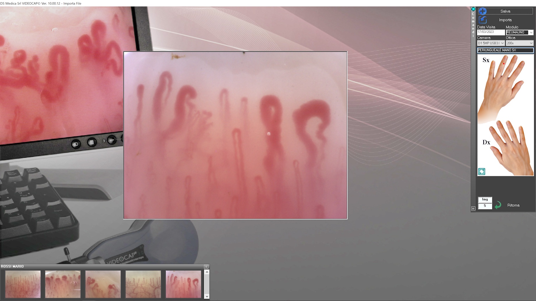
Possibility of capture images by positioning the probe on the body area to be examined and focusing on the image. The “in vivo” images will appear on the screen.
The software is enabled for video recording on hard disk or VHS.
Use of the most sophisticated measurement and image analysis tools
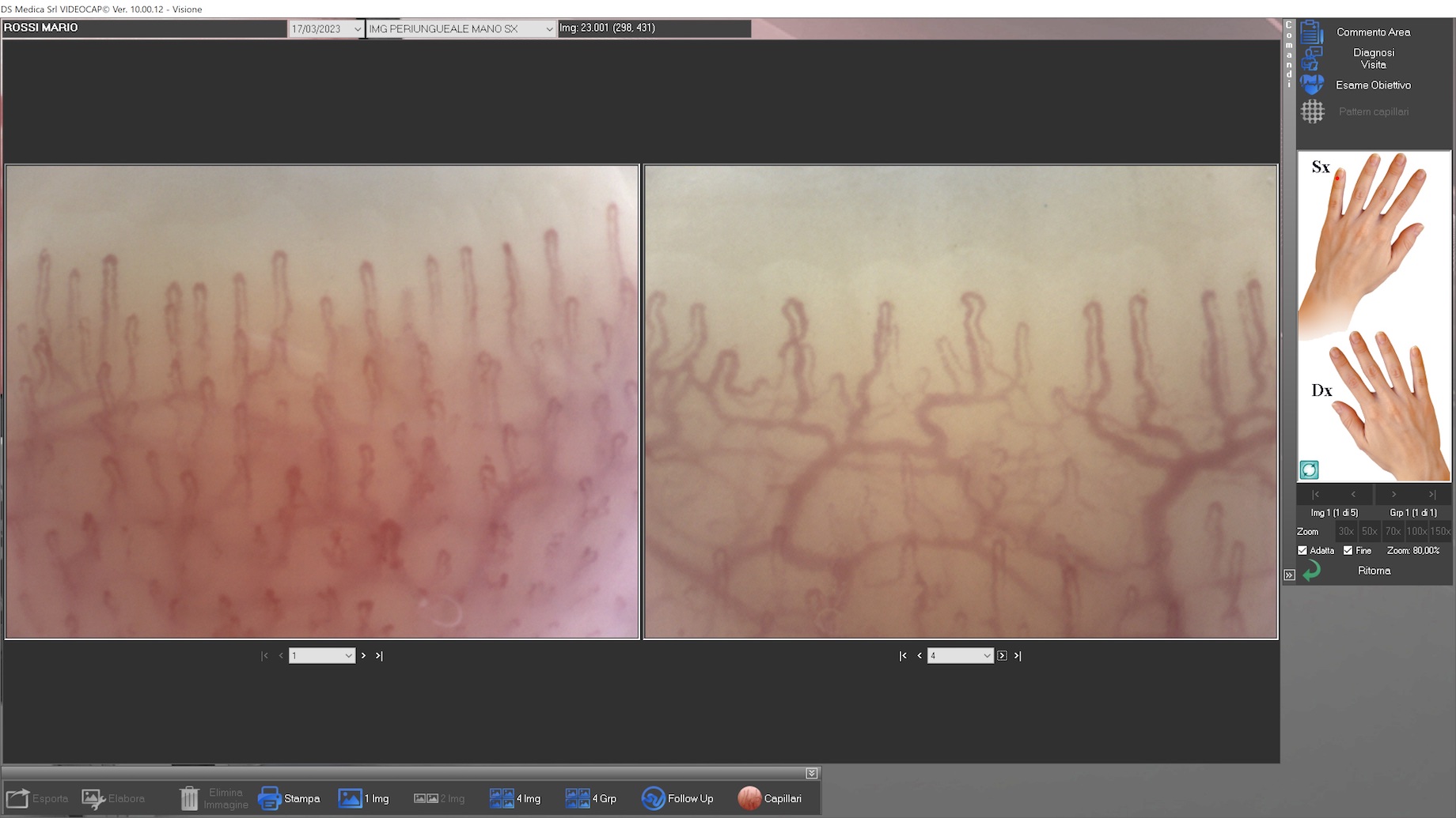
Possibility of comparing the images of a patient obtained during different visits, belonging to different fingers or obtained from other sources (databases, CD-ROM, historical archive created by the user).
Possibility of an accurate and proper follow up.
Possibility of correct and complete reporting according to national and international guidelines. Possibility to print different types of reports; 1, 2 and 4-image reports of the same lesion or visit or follow-up to a specific lesion or visit. All reports can be customized according to the user’s needs.
The software is designed for the use of telemedicine which, however, remains the responsibility of the user.
Have you any question on Videocap® software?
Autocapi®
Autocapi® is an optional module of Videocap software for the automatic capillary counting integrated.
One of the basic requirements for the diagnosis of diseases and the verification of the effectiveness of the treatments implemented, is capillary density. Until now this verification was not objective but operator-dependent.
The Videocap® software is the first software to have an automatic capillary count which allows objectification of the result: it’s the computer that count the capillaries and it is no longer an action performed manually by the operator. This optional function is very important in the field of studies and research because it allows to standardize the result obtained.
Moreover, it allows you to have feedback on mathematical calculations and standardized algorithms, and not on the subjective interpretation of the image.
This function is essential for the verification of the effectiveness of the therapy implemented as it gives an effective quantification of the capillaries present, with an objective result, and no more subjective.
Clinical validation occurred through the study “Automated assessment of absolute nailfold capillary number on videocapillaroscopic images: Proof of principle and validation in systemic sclerosis”.
The configurations
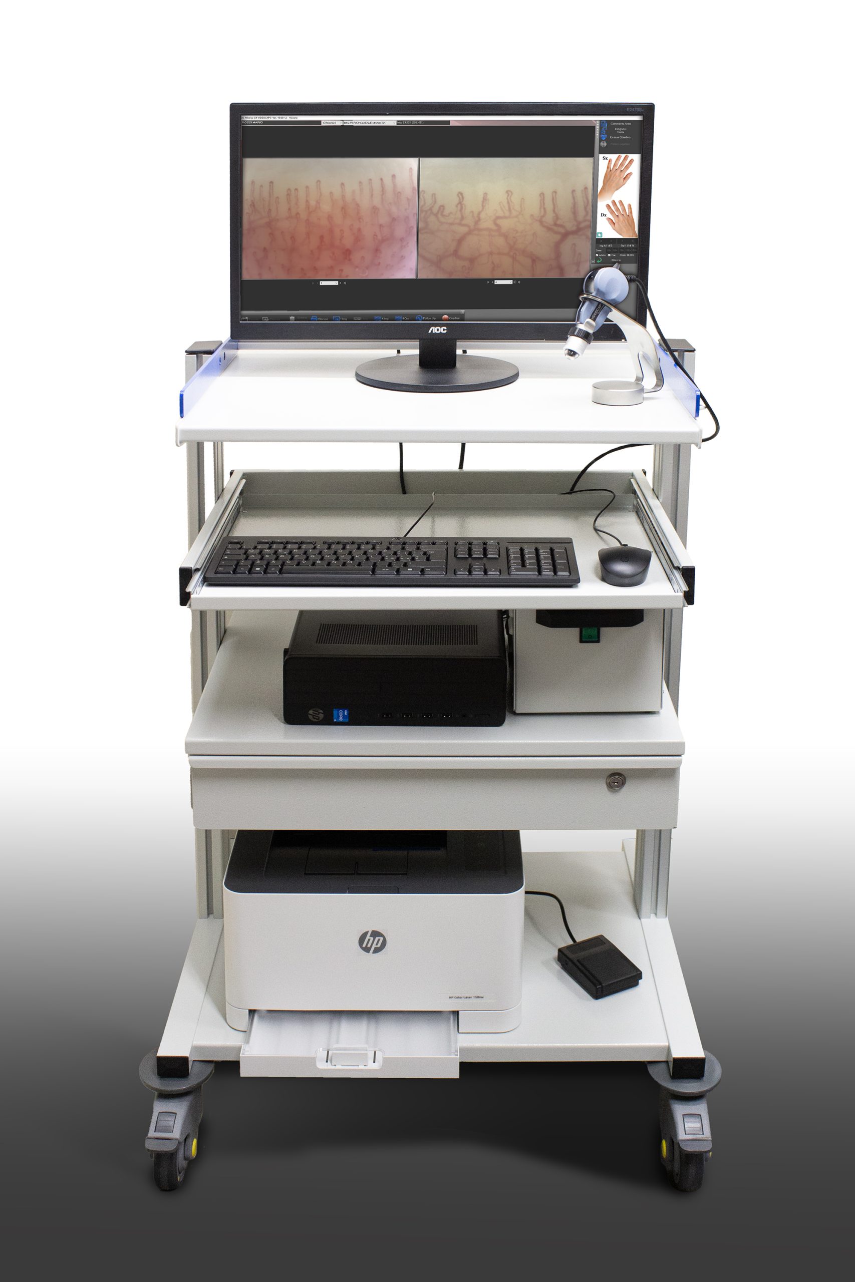
Kit Videocap® Reuma Basic
Computerized workstation for capillaroscopic analysis complete with Videocap D1 3.0
Kit Videocap® Reuma Superior
Computerized workstation for capillaroscopic analysis complete with Videocap D1 3.0, carriage and Autocapy module.
Videocap® is the DS Medica product line dedicated to epiluminescence, immersion and polarized light video biomicroscopes.
DS Medica S.R.L. | Tax ID/VAT 12676030153
Web browsing
Privacy
Customer Support Service
Our customer service to assist in the proper use of software and instrumentation is available Monday through Friday
- © 2023 DS Medica S.r.l.
- Credits